
The best way to diagnose this condition is with a quick visit to the your doctor for a physical examination of your knee. There are many conditions other than meniscus injuries that can cause knee pain such as an ACL tear, patellar or quadriceps tendinitis, a fracture, arthritis or knee bursitis. Getting a proper diagnosis is important so you can treat your condition correctly. This step of knowing what type of meniscus injury you are suffering from is key to your recovery!
Your doctor or orthopedic surgeon will be able to test whether you have a meniscus injury and then will determine what type you have through a variety of tests. It is more difficult to diagnose a lateral meniscus tear than a medial meniscus tear because of its tear shape and location and it may go unnoticed until it is much larger.
To help your doctor achieve a proper diagnosis, he/she will begin with a medical history about you, your current condition and symptoms. The intensity of your pain, the how long you have had your symptoms and the limitations you are experiencing. Details about what and when it started, and whether or not you have ever had treatments (for this or a similar condition in the past) are very helpful in discovering your injury.
Tears of the meniscus can be divided into two groups - those where there is locking, and those where locking is absent. If your knee joint is locking, the diagnosis of meniscus damage is quite clear, whereas the absence of locking in the knee joint makes meniscus damage more difficult to diagnose.
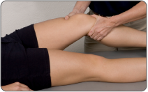
A physical exam will be performed to determine if you have any signs of a meniscus tear or possibly another knee injury. Your doctor will visually assess and palpate (feel) the bones and soft tissue in and around both of your knees to evaluate symmetry and spot any differences. This will identify abnormalities such as inflammation, bone deformity, and atrophied muscles. He/she will press on the injured side of your knee joint to test for point tenderness and help determine the location of your injury or tear. He/she may ask you to complete a series of knee and leg movements such as moving your knee from a straight to bent position (or vice versa), or rotating your knee to see what motions cause pain, weakness, instability and/or grinding, catching, popping or locking. Your knee will also be inspected for fluid, swelling and warmth.

It is important to note that no one single test is sufficient enough to establish 100% accurate diagnosis of meniscus damage. It is also known that test results are not as reliable on occasions where tears are degenerative and/or where there is also additional damage in the form to ligament tears (ie. ACL, MCL).
A McMurray's Test is performed while you lie flat on your back. The doctor will hold your knee with one hand and your ankle in the other. He/she will lift your knee slightly while flexing it to a 45 degree angle. The doctor will feel the medial joint line while pulling your leg toward him/her and rotating your knee. The knee is then brought from full flexion (straight) to a 90 degree angle. If you feel a click during this movement the test is positive; you have a meniscus injury.
A test for joint line tendernessis simply performed by applying pressure over the meniscus area while you are laying flat on your back. If pain occurs, the test is considered positive.
An Apley Grind Test would signifiy a positive result if pain is felt while the knee is flexed to 90 degrees and the leg is rotated both inward and outward. Further to this, the lower leg is pulled away from the knee joint (distraction) and pushed toward the knee joint (compression). If pain flares during compression, it is considered to suggest there exists a meniscal injury.
An Ege's test is performed while you are standing and putting weight on your knees. You begin by standing with your feet 8-10 inches apart. To test the lateral meniscus the doctor will ask you to turn your feet and knees in as far as possible; for medial meniscus tests, you turn your feet and legs out fully. In the appropriate position, you will then squat down and stand up slowly. If you experience pain and/or a clicking sensation (you may even hear it) when your knees are at approximately 90 degrees, the test is considered positive and a meniscus tear is suspected.
A Thessaly Test is performed as follows.. The patient stands on one leg, flatfooted with the knee slightly bent (at 5 degrees) while holding an object (or the examiner) to maintain balance. The patient would then rotate both inward and outward three times, exerting rotational pressure on the knee joint. The patient would then repeat this rotation with the knee bent further (at 20 degrees). The Thessaly Test is considered positive if there is mechanical symptoms (ie. popping, latching, etc) and/or lateral or medial joint line discomfort (pain manifesting horizontally along the seam of the knee joint - lateral = outer knee, medial = inner knee).
A medical professional will often recommend diagnostic testing to obtain more detailed information, and assess the amount and/or type of damage done to your knee and meniscus. There are a variety of different tests available to help them analyze the situation; and the recommendation will be dependent on your injury.
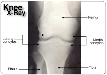
X-rays will provide an image of the overall structure of your knee It is helpful in identifying abnormal bone shapes, fractures, arthritis, and degeneration (wear and tear) within the joint. It can identify a discoid meniscus, or loose bones and bone abnormalities that may mimic a torn meniscus.
CAT scans (or CT - computerized tomography) can be used to provide a 3-dimensional assessment of the bones and soft tissues in and around your knee joint and may be used to identify a meniscus tear.
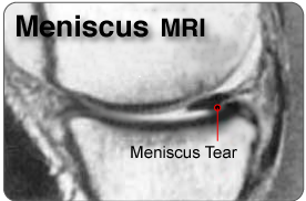
MRIs (magnetic resonance imaging) will provide more detailed information and will help to evaluate the soft tissues in and around your knee joint (muscles, tendons, ligaments, menisci, other connective tissues). It can identify ligament and meniscal damage, and help to determine the extent of your injury, the displacement and degree of your tear, fluid on your knee, a discoid meniscus, ACL or MCL tear, and/or other associated conditions.
The type of diagnostic test recommended will depend on your symptoms and the opinion of your medical professional. Other methods such as electromyogram or arthroscopic surgery can be used to determine the degree and location of your meniscus tear if required.
If you've taken the step to get a proper diagnosis, then you've seen your doctor and may have your MRI results in hand. When it comes to meniscus tears there's a lot of medical terms that might mean very little to you but will actually describe the type of tear you have by using:
The severity of your meniscus tear will vary depending on which meniscus is injured and the location, type and shape of the tear. Such information will be very helpful in your process of recovery, helping you and your doctor decide whether or not surgery may be required and/or if a meniscectomy, repair or knee replacement is advised. The most common locations for meniscus injury are in the posterior horn (back of knee) and the most common type of tear is a longitudinal tear. (source: Campbell's Operative Orthopaedics)
Blood supply to the injured meniscus is critical to healing! The zone of the tear on the meniscus (the blood supply zone it's located in) will influence your ability to heal without surgery. The meniscus tissue is different 'fibrocartilage' than any other soft tissue in the body and has limited blood flow. This can make it difficult for the body to heal a meniscus tear on its own.
Tears in the red zone have the best chance of healing because they have more access to blood supply.
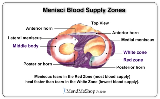
Your tear location is termed red-on-red if both sides of a meniscus tear are in the red zone (the outer part of your meniscus and knee joint). This means both sides of the tear are in tissue that receive a good supply of bloodflow; as a result it experiences a quick healing rate.
Your tear location is termed red-on-white if the tear spans between the red zone and the middle (includes the outside rim and center portion of your meniscus). This type of meniscus tear heals slowly, as the outer edge of the tear generally receives good blood supply whereas blood supply to the inner part of the tear is not as good. Depending on the severity, shape/pattern and size of your tear, this may need surgery. Surgeons will often try to suture (repair) a meniscus when a tear occurs in this zone. This is because they will have determined that there's enough blood supply available to assist with healing after the surgery. When using conservative treatment options, a red-on-white meniscus tear will heal faster than white-on-white tears of the same grade.
Your tear location is termed white-on-white if the tear is in the white zone (the inner part of your meniscus). Tears in this location have a poor healing rate and in most cases they won't heal naturally because there is little to no blood supply. A bucket handle or parrot beak tear (where there is displaced tissue causing locking/catching/clicking in the knee) in the white-on-white zone is usually removed surgically (either through a partial meniscectomy or full meniscectomy) as healing is very unlikely.
Your knee joint can also be divided into the anterior horn (mobile, curved portion at front of meniscus), the posterior horn (less mobile, curved portion at back of the meniscus) and the body (middle section of the meniscus, thicker on outside and thinner on the inside).
There are 3 types of meniscal tears you can experience in these locations:
A partial meniscus tear (partial thickness in depth - meniscus still remain attached) tends to be smaller and more stable because it stays connected to the front and back of your knee and doesn't move about freely. Depending on the location, a partial tear can heal well with non-invasive methods.
A complete meniscus tear (full thickness - tissue separates from your meniscus and tear goes all the way through) tends to be larger and less stable because it hangs by a thread of cartilage. The torn part moves about in your joint which can lead to further complications and damage if not treated.
Degenerative meniscus tears have frayed edges on the inner rim, where the meniscus is thinnest, which can eventually tear in multiple directions and can lead to a completely degenerated meniscus. A piece in the body of the meniscus often moves about in your joint.
The pattern of your meniscus tear will influence your doctor's method of treating your injury. Some patterns are capable of healing themselves through conservative treatments, while others will require surgery to treat severe symptoms (locking of your knee). The shape of your tear will also determine the most appropriate surgical procedure to fix your torn meniscus.
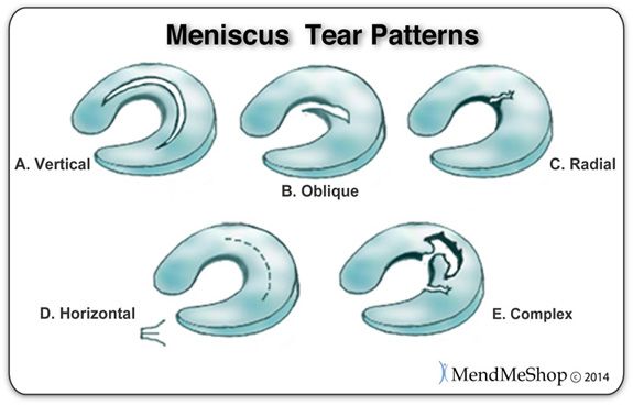
Diagram of meniscal tear patterns:
(A) Vertical or longitudinal (Bucket-handle).
(B) Oblique - tears are known as flap or parrot beak tears.
(C) Radial meniscus tear (or Transverse) - start on the inside white zone and goes to the middle body. A radial meniscus tear can continue to tear and reach the outside red zone. If this happens your surgeon will have no choice but completely remove your meniscus (meniscectomy).
(D) Horizontal cleavage tears - start on the inside white zone and goes to the middle body the meniscus may show signs of degeneration.
(E) Complex degenerative meniscus tears.
Only about 10 - 15% of meniscus tears are repairable, and in most of those cases the meniscus is repaired along with other tissue in the knee (such as the anterior cruciate ligament or ACL). (source: http://www.ckynde.dk/resources/ArtroskopiGenerel.pdf [Campbell's operative orthopaedics, Arthroscopy of the lower extremity] accessed 2019)
The shape of your meniscus tear is important because it will help determine the type of treatment you receive; some tears will heal without surgery, some can be treated surgically and some can't be fixed. Tears come in many shapes and sizes, however there are 3 basic shapes for all meniscal tears: longitudinal, horizontal, and radial. If these tears are not treated, they may become more damaged and develop a displaced tear often referred to as a bucket handle tear (longitudinal), flap tear (horizontal) or parrot beak tear (radial). Complex tears are a combination of two or more of these basic shapes with damage occurring in more than one direction and depth.
A longitudinal tear extends lengthwise, following the collagen fibers that run parallel to the contour of the meniscus. This tear does not go all the way through the meniscus and it divides your meniscus into an inner and outer section; however the tear generally never touches the outer rim of the meniscus. It tends to be more medial than lateral and results from repeated movements. It generally starts as a partial tear in the posterior horn, which can sometimes heal on its own.
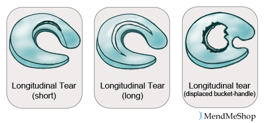
If a longitudinal tear doesn't heal properly it can lead to a displaced tear, known as a bucket handle tear. This is a complete tear that goes all the way through and never touches the inner rim of your meniscus. There is a risk that the handle may flip over and can catch on the femur, locking the joint and increasing pain. This tear accounts for 10% of all meniscus tears, and causes your knee to lock in flexion. It is seen most often in young athletes, and happens in conjunction with 50% of ACL injuries.
The most common location of an injury in your knee is the posterior horn of the meniscus, and longitudinal tears are the most common shape of tears. Small tears limited to the posterior horn of the meniscus won't lock your knee (like other, more severe, tears) but will cause pain, constant / on-going swelling and leave you feeling weak or unstable in the knee. Vertical tears of the meniscus start from either the top or bottom of the meniscus and can be separated into 2 distinct shapes: longitudinal tears and radial tears.
A horizontal tear starts as a horizontal split deep in your meniscus. This tear divides your meniscus into a top and bottom section (like a sliced bun). It is often not visible and moves from the posterior horn or mid section to the inside of your meniscus. Horizontal tears are rare and often start after a minor injury from rotation or degeneration. It occurs most often in your lateral meniscus but however it is noted in both menisci. Horizontal meniscus tears are degenerative in nature. They are more likely to occur in people as they age who may or may not already show signs of having osteoarthritis.

A complete horizontal tear resulting in displaced tissue is also referred to as a horizontal flap tear and can develop if your tear is overlooked or left alone. This type of tear is horizontal on the surface of your meniscus and creates a flap that flicks when your knee moves. It is a result of a strong force that tears your meniscus from the inner rim; it can easily become a complex tear if left untreated. Often, the flap is trimmed away during surgery to prevent further tearing. Since the periphery of the meniscus is not compromised and there is enough tissue left to heal, the cushioning function of the meniscus is maintained. If this tear extends from the apex of your meniscus to the outer rim, you may develop a meniscal cyst (a mass that develops from a collection of synovial fluid along the outside rim of the meniscus) and/or experience increased swelling in and around the knee.
A horizontal tear is a tear that is most commonly amenable to meniscus repair. Rather than removing the damaged portion of the meniscus, a horizontal tear may be able to be sewn together though healing will depend on adequate bloodflow in the area.
A radial tear starts as a sharp split along the inner edge of your meniscus and eventually runs part way or all the way through your meniscus, dividing it into a front and back section (across the middle body instead of down the length). This tear generally occurs between the posterior horn and middle section and is seen frequently in your lateral meniscus.
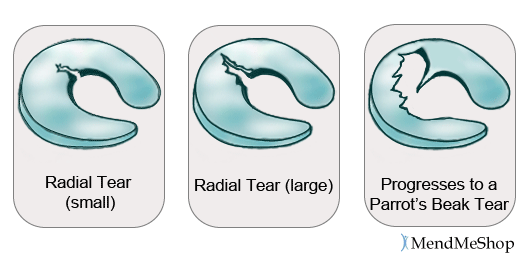
A small tear is difficult to notice, but when a radial meniscus tear grows and becomes a complete tear it will open up (meaning some of the meniscus gets displaced) and look like a part is missing. A displaced radial tear is called a Parrot's Beak tear as the tear generally resembles the curved shape of a parrots beak. Parrots beak tears have been found to typically occur in the thicker portion of your lateral meniscus. As it gets larger, it will catch or lock more frequently, and prevent your meniscus from protecting the articular cartilage during weight bearing. This tear is usually the result of a traumatic event or forceful and repetitive stress activities and it is often associated with other injuries such as ACL tears. Young athletes tend to suffer from combination tears called radial/parrot beak tears (the meniscus splits in 2 directions).
Similar to longitudinal tears, radial tears usually occur due to some kind of acute trauma and appear in younger and more physically active people. If you have undergone partial meniscectomy surgery, you are more likely to experience a radial tear. This may happen due to changes in knee movement and function because of the previous removal of meniscus tissue. Radial tears can be more damaging to your knee than longitudinal tears as radial tears affect the ability of your meniscus to transfer load and bear weight on your knee.
The size of your meniscus tear will have some affect on your ability to heal the tear through conservative treatments. Meniscus tears under 1 cm can heal without surgery if it's located in the red-red or red-white zone (with some blood supply for healing). Tears that are 1.5 cm to 4 cm usually require surgery. The size of the tear becomes important because if the tear in the tissue is too large it will not heal without the help of your surgeon.
To understand why the size of your tear is important, let's imagine you have 2 slices of bread in front of you. You tear one just a little (less than an inch) and place it down. When you do this the bread will naturally pull the edges of the small tear back together. You tear the 2nd slice of bread in half (or more). When you place the bread down it will be floppy, and remain out of shape. You would need your hands, some peanut butter, or something else to force/keep both sides of the tear together. Your meniscus is no different, small tears have the ability to heal since the edges are still close enough to heal back together whereas large tears need sutures to bring the two sections of the tear back together.
Your age will directly influence the causation of your meniscus injury - the younger you are, the more likely a torn meniscus will occur during a sport, whereas torn menisci are more likely occur through degenerative isues as you age. It's been proven that the meniscus becomes weaker over time resulting in degenerative meniscal tears; 60% of individuals over the age of 65 will experience a degenerative meniscus tear. (reference: 1)
A degenerative tear would typically require a partial meniscectomy to remove damaged and displaced tissue. A meniscal repair isn't possible to fix degenerative damage because of the jagged and torn nature of the meniscus. A meniscal repair of degenerative tissue would be very difficult to perform and reduces the rate of success for healing. Because of this fact, your age increases the likelihood that your surgeon will feel that your tear can't be repaired.
Click HERE to Go To Our Online Store We take all major credit cards and Paypal.
If you have questions, call our office at 1-866-237-9608 (toll free continental US).
We are currently offering FREE SHIPPING and a 60 day trial period on all our Wraps.
Product specialists are available 9:00 am to 5:00 pm Eastern Standard Time Monday to Friday.
If any question or concern arises, call us or simply send us an email at any time (we check our emails constantly all throughout the day and night.. even on holidays!). We will respond as soon as possible.
I want to learn more about Post-Surgery Recovery
I want to learn all about Types, Patterns, Shapes & Severity of Meniscus Tears
I want to learn more about TShellz Wrap® Circulatory Boost
I want to learn more about Ice & Heat: Which Is Better For Treatment?
I want to learn more about Meniscus Treatments
I want to learn more about different types of Meniscus Surgery
During your recovery, you will probably have to modify and/or eliminate any activities that cause pain or discomfort at the location of your soft tissue injury until the pain and inflammation settle. The more diligent you are with your treatment and rehabilitation, the faster you will see successful results!
Please be aware that this information is neither intended nor implied to be a substitute for professional medical advice. CALL YOUR HEALTHCARE PROVIDER IMMEDIATELY IF YOU THINK YOU MAY HAVE A MEDICAL EMERGENCY. Always seek the advice of your physician or other qualified health provider before using any of our outstanding products to make sure they are right for you and your condition or if you have any questions regarding a medical condition. Always see your doctor for a proper diagnosis as there are often many injuries and conditions (some very serious) that could be the cause of your pain.
© 2025 In.Genu Design Group, Inc. Contact Us